Contact us
Request for: consultancy, collaboration, amending booking rules, account and training
Downloads
User regulations, booking rules, charging scheme, course material, software, protocols and more
Request for: consultancy, collaboration, amending booking rules, account and training
User regulations, booking rules, charging scheme, course material, software, protocols and more
Dear Users,
Thank you so much for joining the celebration for 1000 UMIF users which was held on 01.03.2024.
Despite the strike on the same day, there are over 60 people joined the party. The 1000# user got a small prize from the UMIF team and afterwards we had our international buffet, good music and relaxing time.
I hope you all enjoyed the event and here I shared some images from the night. Thank you for being a part of it and looking forward to seeing you next time!


best wishes
Antonio Virgilio Failla
Dear Users
from April 16 - 18, 2024, there will be a super interesting workshop from one of the world leading microscope manufacturer Abbelight at CSSB with a focus on DNA-PAINT.
DNA-PAINT is a powerful super-resolution light microscopy technique that might be rather easy to use and allowed multiplex imaging. This approach achieves a resolution of a few ten nm (https://www.abbelight.com/dna-paint/).
Please be aware that UMIF has a super resolution microscope of Abbelight!!!!!
If you are interested in this technique, please join an online prep meeting on March 14, 2024 at 16:00. During the prep meeting the technique will be explained and we can discuss experiments that you might want to carry out with your own samples. Abbelight can then provide DNA-PAINT staining reagents that you can use beforehand and then measure your samples at the workshop.
If you want to join the workshop and/or the online prep meeting, please send an email to
Please diffuse this information to anyone might be interested, this is a unique chance to use.
Best wishes
Antonio Virgilio Failla
Dear users,
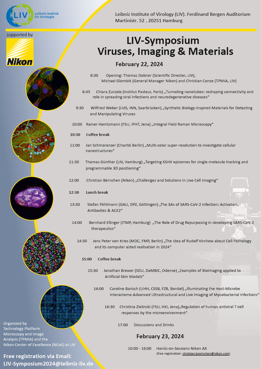
A very Interesting symposium with exceptional guests will take place at LIV (for more information please have a look at the flyer). Registration can be accomplished following the indication on the flyer. Please diffuse this information to every one who might be interested.
Best wishes
Antonio Virgilio Failla
Dear users,
Interherence GmbH is a spin-off of the Max Planck Institute for the Science of Light in Erlangen, Germany. Their product, VAHEAT, is a very interesting compact and powerful device to control the temperature of your sample during live imaging. This device can be customized on any microscope allowing the opportunity to have live cell imaging also in microscopes that does not have a proper incubator (for more information please have a look at the flyer ). Please diffuse this information to anyone who might be interested but not belonging to the imaging facility. If you express your interest in testing the device by emailing me (

Best wishes
Antonio Virgilio Failla
Dear users,
Depending on your interest we might be entitled to test a very important compact setup for in vivo imaging made by Optiscan (for more information please have a look at the flyer). Please consider this setup might be useful also to colleagues working in the hospital and that are not using the imaging facility. Please do not hesitate to diffuse to them this information.
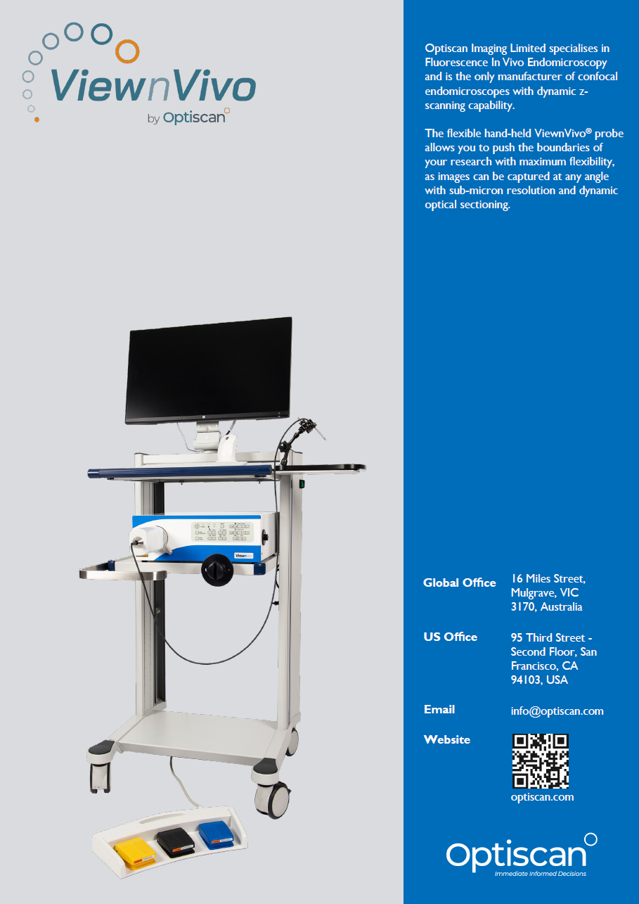
If interest from you arises please contact me (
Best wishes
Antonio Virgilio Failla