Contact us
Request for: consultancy, collaboration, amending booking rules, account and training
Downloads
User regulations, booking rules, charging scheme, course material, software, protocols and more
Request for: consultancy, collaboration, amending booking rules, account and training
User regulations, booking rules, charging scheme, course material, software, protocols and more
Dear users,
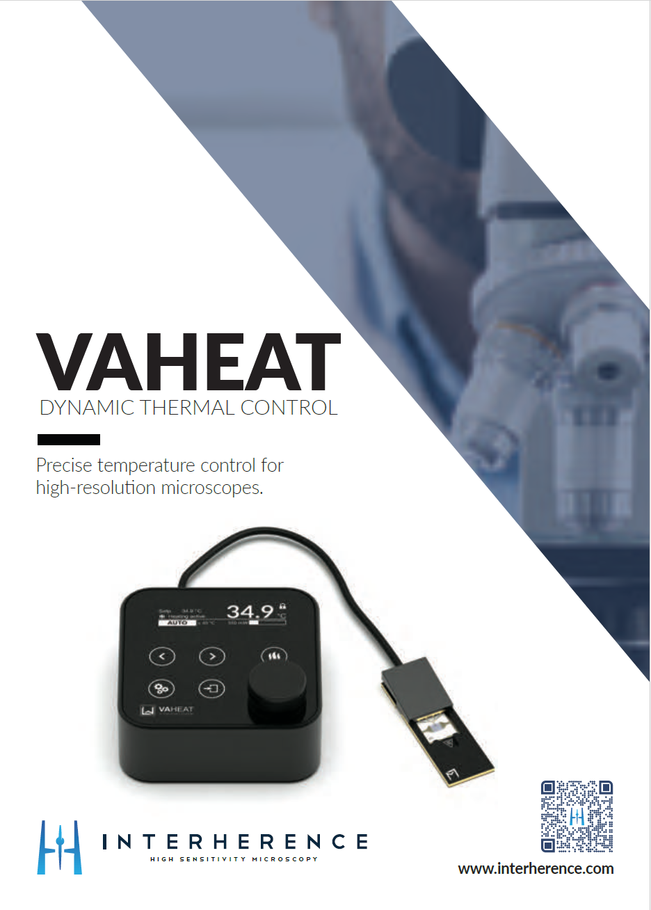
Interherence GmbH is a spin-off of the Max Planck Institute for the Science of Light in Erlangen, Germany. Their product, VAHEAT, is a very interesting compact and powerful device to control the temperature of your sample during live imaging. This device can be customized on any microscope allowing the opportunity to have live cell imaging also in microscopes that does not have a proper incubator (for more information please have a look at the flyer ). Please diffuse this information to anyone who might be interested but not belonging to the imaging facility. If you express your interest in testing the device by emailing me (
Best wishes
Antonio Virgilio Failla
Dear users,
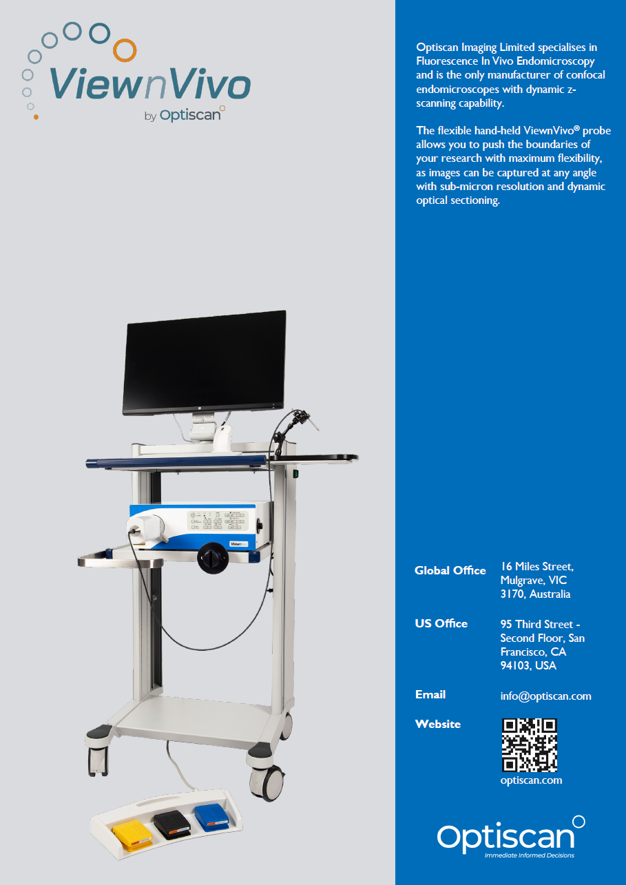
Depending on your interest we might be entitled to test a very important compact setup for in vivo imaging made by Optiscan (for more information please have a look at the flyer). Please consider this setup might be useful also to colleagues working in the hospital and that are not using the imaging facility. Please do not hesitate to diffuse to them this information.
If interest from you arises please contact me (
Best wishes
Antonio Virgilio Failla
Dear users,
Thanks to all of you the light microscopy facility has achieved incredible milestones, personal as well as scientific.
We are aiming to celebrate with all of you one of those: our user number 1000.
The user number 1000 prize will be given on Friday 01.03.2024 in seminar room 14 at 5pm.
The user number 1000 prize will give us the chance to organize a buffet with Italian Chinese, German and maybe also your favorite food.....
In order to organize food and drinks If you wish to participate (and you are all super warmly invited to do so) please register on this link:
https://docs.google.com/spreadsheets/d/1bIfcwO1KOWy__x2AMLd5_G0pjNjzVnJhm0tXA_WxvYo/edit?usp=sharing
As you can see from the registration form if you wish you can join us preparing some food but this is a free willing option
We are looking forward to share with you good food and mood
best wishes
Antonio Virgilio Failla
Sven Hey (Ag Linder) has recently published “KIF16B drives MT1-MMP recycling in macrophages and promotes co-invasion of cancer cells” in “Life Science Alliance” (DOI: 10.26508/lsa.202302158). Beside that we are all warmly invited to read this very interesting work, we take much pride to inform you that one of the images associated to this manuscript has been awarded to be the cover image for the entire issue!!!
Congratulation to Sven that expressed his scientific and imaging talent using UMIF microscopes

About the image: Spheroid of H1299 carcinoma cells expressing GFP (green), embedded in collagen I matrix, together with primary human macrophages; F-actin of both cell types stained by Alexa568-phalloidin (red), nuclei by DAPI (blue); maximum projection of confocal z-stack.
The cover image was acquired by using UMIF´s SP8 microscope.
Dear users.
Our workshop season becomes hot:
The next appointment I would like to bring to your attention is with Evident.
Evident will promote a compact, easy to handle, powerful slide scanner imaging system that is not the classical wide field slide scanner: it can produce images with confocal resolutions also in 3D.
When: 26.09-28.09.
Where: lecture: seminar room 14, building N27
practical sections: seminar room 1, building N45
To get more information and for the registration link please look at the flyer below (registration is free of charge).
As always you are all warmly invited to join and let join everyone might with to participate.
Thank you very much to make this fantastic imaging community
best wishes
Antonio Virgilio Failla
